How it Works
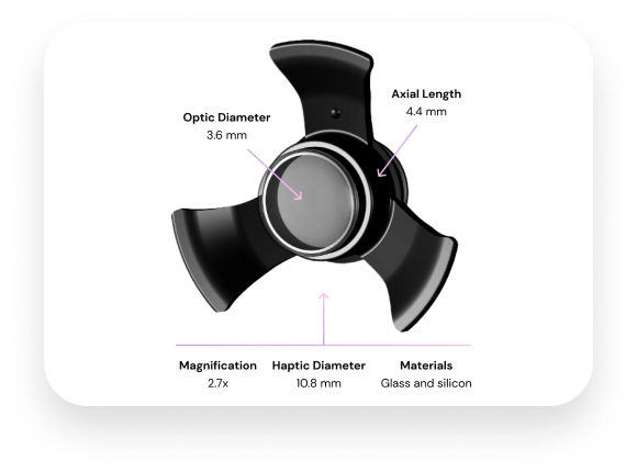
The SING IMT™ has a Galilean design, based on ultra-precision, wide-angle micro-optics that, in combination with the optics of the cornea, create a telephoto system that magnifies objects in view. Implantation inside the eye allows the patient to see using natural eye movements in both stationary and dynamic environments.
The telescope is about the size of a pea (3.6 mm; 4.4 mm length) and is surgically placed inside the eye.
Reducing the Impact of the Blind Spot
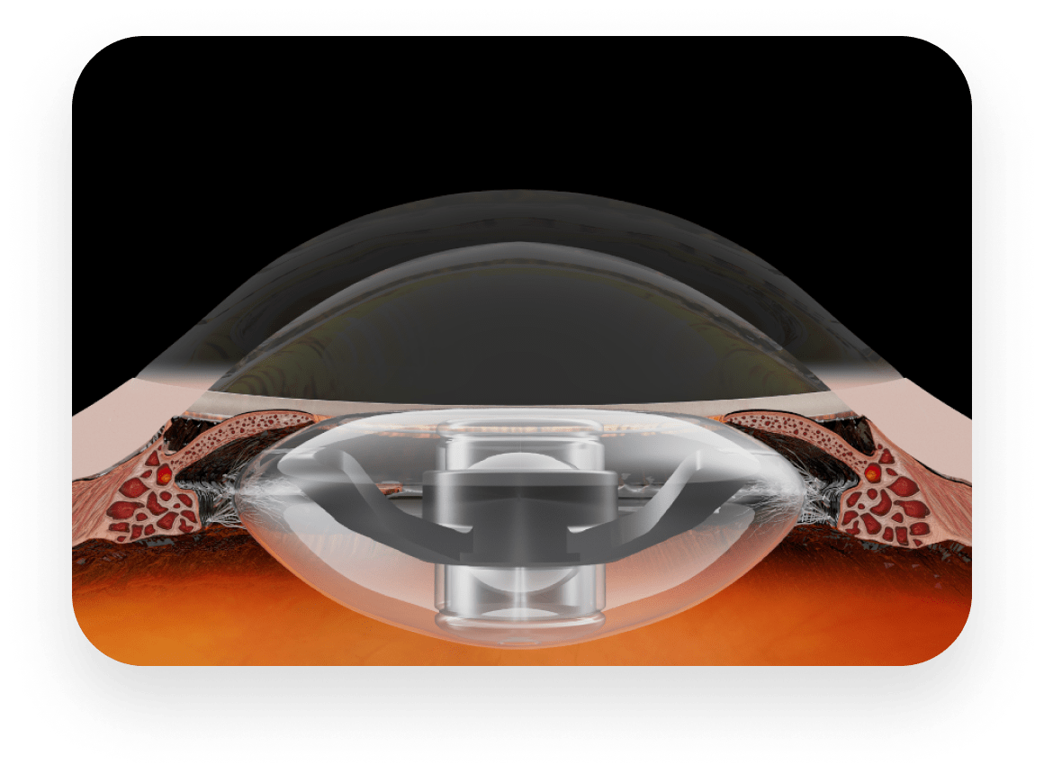
At the time of cataract surgery, the SING IMT is surgically implanted monocularly in the capsular bag after removal of the eye’s lens and is held in position by haptics. The SING IMT, along with the cornea, enlarges images in front of the eye approximately 2.7 times their normal size. The nominal field of view of 20°-24° is projected onto approximately 54° of the retina, effectively reducing the impact of the central scotoma (blind spot) in central vision. The magnification allows central images to be projected onto healthy perimacular areas of the retina instead of the macula alone where breakdown of photoreceptors and loss of vision has occurred.
This magnification and projection helps reduce the ‘blind spot’ and allows the patient to distinguish and discern images that may have been unrecognizable or difficult to see due to the central scotoma associated with their AMD. The central scotoma is not eliminated or cured, but patients can better see “around” it as images are projected onto healthier parts of retina undamaged by AMD.
SING IMT Construction
The SING IMT is housed in a prosthetic device (refer to professional use information) composed of three primary components: a glass capsule that contains wide-angle micro-optical elements; a clear polymethylmethacrylate (PMMA) carrier; and a blue PMMA light restrictor. The sealed optical component is snap-fitted into the carrier plate.
Biocompatibility and MRI Compatibility
The telescope prosthesis uses biocompatible materials for long-term ocular implantation per ISO 10993-1. One of the internal components (not in contact with body fluids or tissue) of the device contains stainless steel, which has been evaluated for MRI compatibility and determined to be MR-Conditional.*
*MRI testing was performed to evaluate the magnetic field interactions, heating, and artifacts of the telescope prosthesis induced by a commercial 3-Tesla MRI system. Based on the MRI testing information, the telescope prosthesis will not present any additional hazard or risk to a patient undergoing an MRI procedure using a scanner operating with a static magnetic field of 3-Tesla or less and under the MRI-related heating conditions used for the evaluation (MRI for 15 minutes at an MR system reported whole body averaged specific absorption rate, SAR, value of 3 W/Kg). The MRI image quality may be compromised if the area of interest is the same as or close to the position of the device (in this case, it may be necessary to optimize MR imaging parameters to compensate for the presence of the implant).